Many gym enthusiasts claim that rapidly growing skin is the reason they develop stretch marks. However, as readers of The Stretch Mark Secret know, that explanation is incomplete. The real issue goes much deeper and involves a combination of hormonal imbalance and fibroblast dysfunction.
While overstretched skin may contribute slightly, the primary reason why many bodybuilders and athletes experience stretch marks lies in the activity of fibroblasts, the connective tissue cells responsible for producing collagen and elastin. These fibroblasts can malfunction due to disrupted hormonal signaling, leading to weak or disorganized connective tissue that tears under stress.
The Different Types of Skin Cells
Before understanding why fibroblasts can become dysfunctional, it helps to know that the skin contains several types of cells:
- Keratinocytes
- Melanocytes
- Langerhans cells
- Merkel cells
- Dermal fibroblasts
Dermal fibroblasts play a crucial role because they produce collagen and elastin, which maintain the structure and elasticity of the skin. When fibroblasts stop functioning correctly, the skin loses its ability to stay strong and flexible. This sets the stage for stretch mark development, especially in bodybuilders who undergo rapid muscle growth and hormonal fluctuations.
How Hormones Affect Fibroblast Function
Fibroblasts are found throughout the entire human body, not just in the skin. They adapt to the needs of different tissues depending on the surrounding hormonal environment. Research shows that fibroblasts have numerous receptors for sex hormones, such as testosterone and estrogen. (1, 2, 3, 4, 5)
Let’s do a little detour into all the different scenarios of fibroblast dysfunction caused by sex hormone imbalances. After that, you will understand why stretch marks aren’t caused by overstretched skin but rather by sex hormone imbalances that lead to malfunctioning fibroblasts, which in turn produce excessive amounts of collagen-rich scar tissue instead of smooth skin.
Example: The Vocal Folds
The vocal fold is primarily made up of collagen, which is produced by fibroblasts. (6)
During puberty, sex hormones stimulate fibroblasts in the vocal fold to make more collagen and grow the vocal cords in both boys and girls. (7,8) The voice change is more pronounced in boys than in girls, but girls also go through a change of vocal tonality during puberty.
It’s been shown that the combined administration of testosterone, estradiol, and corticosterone suppresses fibroblast activity in the vocal fold, but testosterone alone increases fibroblast activity. (9)
This demonstrates how complex and sensitive fibroblast regulation is. A delicate hormonal balance is necessary for optimal tissue health.
Example: Fibroblasts in the Heart Muscle
Fibroblasts are also active in the heart muscle, and their activity is heavily influenced by testosterone. Excessive steroid use can overstimulate fibroblasts, leading to an overproduction of collagen relative to elastin. This imbalance stiffens arterial walls and raises the risk of heart disease.
Studies show that low testosterone can also increase fibroblast activity and elevate cardiovascular risk. (10)
Conversely, too much testosterone raises fibroblast activity as well. (11, 12)
The conclusion is that both too much and too little testosterone can lead to an increase in fibroblast activity and make them produce too much collagen.
Fibroblasts and Hormone Conversion
The relationship between hormones and fibroblasts goes even deeper. Research has shown that fibroblasts can be converted into Leydig-like cells, which are very similar to real Leydig cells, the testosterone-producing cells in the testicles. (13)
These converted fibroblasts were even capable of producing testosterone themselves. Yes, you read that correctly. Simple collagen-producing fibroblasts can produce testosterone. This is really important to understand the connection between scar tissue (stretch marks) and sex hormone imbalances.
The female reproductive system also contains many fibroblasts. Uterine and fallopian fibroids are, as the name implies, the result of dysregulated fibroblast activity. Uterine/fallopian fibroids contain excessively large amounts of collagen. (14, 15)
These fibroids often arise from prolonged estrogen dominance, either from internal imbalance or environmental xenoestrogens. (16)
Up to 80% of all women develop fibroids during their lifetime. That’s why, just like men, women should avoid sources of xenoestrogens (e.g. from plastic bottles) and high amounts of phytoestrogens (e.g. by eating too many soy products). Men already know they shouldn’t be soy boys. The same goes for the ladies: don’t be a soy girl.
Fibroblasts and Cancer
Fibroblast dysfunction is not only involved in stretch marks or fibroids but also plays a role in several types of cancer. In breast cancer, fibroblasts are the dominant cell type driving tumor growth and metastasis. (17, 18)
Similar processes occur in other cancers where fibroblasts promote abnormal tissue expansion. (19)
Again, hormone-like substances in the modern environment contribute to this dysfunction. This highlights how sensitive fibroblasts are to hormonal signals and why balancing hormones is essential for both health and appearance.
Testosterone, Collagen, and Skin Health
The effects of testosterone on collagen production vary depending on context. Some studies show that testosterone stimulates fibroblast activity and collagen synthesis (20), while others show the opposite (21).
This mirrors other paradoxical hormonal effects, such as dihydrotestosterone (DHT), which can promote hair growth in one area of the body while causing hair loss on the scalp (22). Men with high DHT levels experience an increase in body hair growth as well as a decrease in hair growth on their head (male-pattern baldness). These contradictions underscore that hormone balance, not absolute levels, determines whether fibroblasts work properly or malfunction.
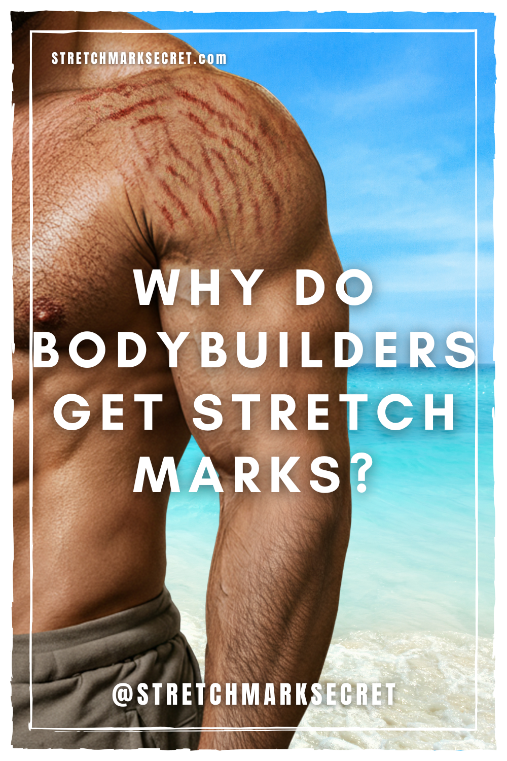
Why Stretch Marks Appear in Specific Areas
Men usually develop stretch marks around the shoulders, chest, and arms, while women tend to develop them around the hips, thighs, and breasts. This pattern reflects the distribution of steroid hormone receptors in the body. (23)
Men have more receptors in their upper body, where testosterone activity is strongest. Women have more receptors in their lower body, influenced by estrogen. These areas correspond exactly with where stretch marks commonly appear in each sex.
This receptor distribution explains why both men and women get stretch marks in predictable locations: men around their shoulders and glutes, women around their glutes and breasts. It is not because the skin was simply stretched too fast, but because fibroblast dysfunction in those hormone-sensitive regions weakens connective tissue integrity and makes the fibroblasts produce too much scar tissue (stretch marks) instead of flawless skin.
Correlation Is Not Causation
Muscle growth and stretch marks often appear together, but that does not mean one directly causes the other. Hormonal changes that drive muscle growth also influence fibroblasts, which explains why both phenomena occur at the same time at the same location.
Stretch marks are a sign that the skin’s connective tissue has been compromised at the cellular level. This problem cannot be fixed by moisturizing alone. Instead, it requires addressing the internal hormonal imbalances that impair fibroblast function.
Conclusion
Stretch marks in bodybuilders are not caused by skin stretching too fast. They are the result of hormonal disturbances that disrupt fibroblast activity. Steroid abuse, poor diet, and chronic stress can all lead to these imbalances.
To restore skin integrity, you need to correct the underlying causes by improving hormonal health, nutrient intake, and fibroblast function.
If you want to understand these processes in more detail and learn how to reverse stretch marks naturally, download a free preview chapter of The Stretch Mark Secret using the link below.
References
(1) https://www.ncbi.nlm.nih.gov/pmc/articles/PMC3763909/
(2) https://pubmed.ncbi.nlm.nih.gov/17326004/
(3) https://pubmed.ncbi.nlm.nih.gov/32550699/
(4) https://www.sciencedirect.com/science/article/abs/pii/S0303720717304926
(5) https://www.sciencedirect.com/science/article/pii/S0022202X9190102V
(6) https://onlinelibrary.wiley.com/doi/full/10.1002/lio2.993
(7) https://www.ncbi.nlm.nih.gov/pmc/articles/PMC4330318/
(8) https://journals.sagepub.com/doi/abs/10.1177/000348940010901112?journalCode=aora
(9) https://academic.oup.com/endo/article/156/3/1000/2423120
(10) https://pubmed.ncbi.nlm.nih.gov/25125004/
(11) https://pubmed.ncbi.nlm.nih.gov/25125004/
(12) https://www.ncbi.nlm.nih.gov/pmc/articles/PMC7925058/
(13) https://www.sciencedirect.com/science/article/pii/S2213671120302459
(14) https://www.ncbi.nlm.nih.gov/pmc/articles/PMC6488189/
(15) https://clinicaltrials.gov/ct2/show/NCT03444987
(16) https://pubmed.ncbi.nlm.nih.gov/24316382/
(17) https://www.sciencedirect.com/science/article/abs/pii/S1084952120301695
(18) https://www.ncbi.nlm.nih.gov/pmc/articles/PMC7352995/
(19) https://www.frontiersin.org/articles/10.3389/fimmu.2019.01835/full
(20) https://www.ncbi.nlm.nih.gov/pmc/articles/PMC4330318/
(21) https://www.jstage.jst.go.jp/article/endocrj/68/3/68_EJ20-0344/_html/-char/en
(22) https://www.ncbi.nlm.nih.gov/pmc/articles/PMC4171668/
(23) https://www.hindawi.com/journals/jos/2011/702735/